
PROTOPLASM
- The living contents of a cell are termed as protoplasm.
- Protoplasm consists of:
- Cytoplasm
- Nucleus
CYTOPLASM:
Cytoplasm is the protoplasm of a cell outside the nucleus.
COMPONENTS OF CYTOPLASM:
Cytoplasm has three main components which are:
- Cytosol
- Organelles
- Cytoplasmic inclusions
CYTOSOL
- The soluble part of cytoplasm is the cytosol and forms the ground substance of cytoplasm.
- Chemically cytosol is 90% water forming a solution containing all the fundamental molecules of life.
- In cytosol, small molecules and ions may form true solutions and some large molecules form colloidal solutions.
- Colloidal solution may be a solution (non-viscous) or a gel (viscous).
ORGANELLES
- Organelles are membrane bound structures found suspended in the cytosol.
- Organelles act as the machinery of the cell performing specific tasks.
- Following are the main organelles found in a cell.
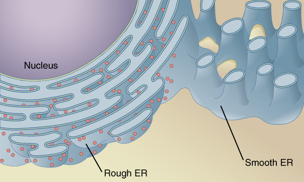
ENDOLASMIC RETICULUM
- Endoplasmic reticulum is a network of membranous channels extending throughout the cytoplasm of a eukaryotic cell.
- These channels are often continuous with the plasma membrane and also appear to be in contact with the nuclear membrane.
STRUCTURE OF ENDOPLASMIC RETICULUM:
- The general structure of endoplasmic reticulum consists of a network of interconnected flattened, membrane bound sacs called cisternae.
- Cisternae is the basic structural unit of endoplasmic reticulum. The sacs are held in shape by the cytoskeleton.
- The phospholipid membrane encloses the cisternal space which is also termed as lumen.
- The lumen is continuous with the perinuclear space but separate from the cytosol.
MORPHOLOGICAL FORMS OF ER:
Endoplasmic reticulum exists in two morphological forms:
- Rough endoplasmic reticulum, which is studded with ribosomes and is involved in protein synthesis.
- Smooth endoplasmic reticulum, which lack attached ribosomes and does not take part in protein synthesis.
FUNCTIONS OF ENDOPLASMIC RETICULUM:
ROUGH ENDOPLASMIC RETICULUM:
RER is involved in protein synthesis. After synthesis the proteins are either stored in the cytoplasm or transported out of the cell through these channels.
SMOOTH ENDOPLASMIC RETICULUM:
SER is involved in several processes which are as follows:
- SER helps in the metabolism of different types of molecules, especially lipids.
- SER brings about the detoxification of harmful drugs.
- SER is responsible for the transmission of impulses such as in nerve and muscle cells.
- SER plays an important role in the transport of materials from one part of the cell to the other.
- SER also provides mechanical support to the cell for maintaining its shape.
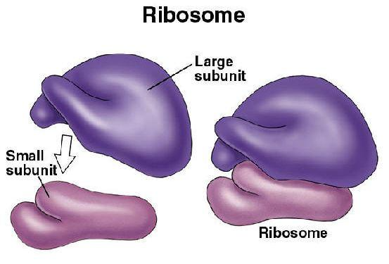
RIBOSOMES
Ribosomes are tiny granular structures present in both prokaryotic and eukaryotic cells.
DISCOVERY: Ribosomes were first studied by Palade in 1955.
OCCURRENCE: Ribosomes may be present within a cell in two forms:
- Attached with RER as tiny granules.
- Freely dispersed in the cytoplasm.
COMPOSITION:
- Ribosome is composed of RNA and proteins therefore they are also termed as ribonucleic-protein particles.
- The RNA present in ribosomes is called ribosomal RNA.
SYNTHESIS:
New ribosomes are assembled in the nucleolus of the nucleus from where they are transported into the cytoplasm via nuclear pores.
SUBUNITS OF RIBOSOMES:
Ribosomes are mainly the combination of two subunits:
- Large subunit
- Small subunit.
The two subunits attach with each other in the presence of Mg2+ ions leading to the formation of a ribosome.
PROKARYOTIC AND EUKARYOTIC RIBOSOME:
Based on the differences in the subunits, ribosomes may be of two types:
Prokaryotic ribosome:
- It is also termed as the 70s ribosome (S= Svedberg unit used in ultracentrifugation).
- Prokaryotic ribosomes result from the combination of 50s large subunit and a 30s small subunit.
Eukaryotic ribosome:
- It is also known as the 80s ribosome.
- Eukaryotic ribosome results from the combination of 60s large subunit and 40s small subunit.
FUNCTIONS OF RIBOSOMES:
- Ribosomes are involved in the synthesis of proteins.
- Ribosomes bring about linkage of amino-acids in a specified order leading to the production of polypeptide chains in the translation phase of protein synthesis.
POLYSOMES:
A group of ribosomes attached to messenger RNA is termed as polysomes.
GOLGI APPARATUS
Golgi apparatus is a complex system of interconnected tubules found virtually in all eukaryotic cells.
DISCOVERY: Golgi apparatus was discovered by Camillo Golgi in 1898.
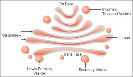
STRUCTURE:
- Golgi apparatus consist of stacks of flattened, membrane bound sac called cisternae.
- These cisternae along with associated vesicles form the Golgi complex.
- Cisternae are continuously formed by the fusion of vesicles which arise by budding off from smooth endoplasmic reticulum.
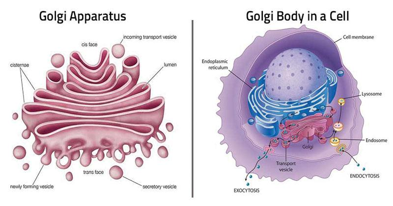
SURFACE OF GOLGI APPARATUS:
Golgi apparatus consists of two ends or surfaces:
- The cis or forming face.
- The trans or maturing face.
Cis Face:
- The convex surface is the cis or forming face.
- It is oriented towards the endoplasmic reticulum.
- Proteins formed in the ER are transferred to the cis face of Golgi apparatus.
Trans Face:
- The concave surface is the trans face.
- The trans face is oriented towards the plasma membrane of the cell
- Secretory vesicles pinch off from the trans face.
FUNCTIONS OF GOLGI APPARATUS:
- Secretions formed within the cell are passed to the outside through the Golgi apparatus where they are modified and packed into vesicles.
- The most important function of this apparatus is to modify the proteins and lipids by adding carbohydrates and converting them into glycoproteins and glycolipids before secretion.
LYSOSOMES
Lyso means splitting while soma means body. Lysosomes are found in most eukaryotic cells.
DISCOVERY: Lysosomes were isolated as a separate component by De Duve in 1949.
OCCURRENCE: Lysosomes are found in abundance in cells exhibiting phagocytic activity such as macrophages.
STRUCTURE:
- Lysosomes are simple sacs bound by a single membrane.
- Lysosomes are rich in hydrolytic enzymes such as acid phosphatase.
FORMATION OF LYSOSOMES
- Lysosomal enzymes are synthesized on RER and further processed in the Golgi apparatus.
- The processed enzymes are budded off as Golgi vesicles and are called as primary lysosomes.
- Primary lysosomes upon fusion with phagocytic vacuole give rise to secondary lysosome.
FUNCTIONS OF LYSOSOMES:
Lysosomes are responsible for the processes of phagocytosis and autophagy in the cells.
Phagocytosis:
- Phagocytosis, also regarded as eating process of cells, is carried out by lysosomes.
- Any foreign object that gains entry to the cell is immediately engulfed by the lysosome and is broken down into digestible pieces by lysosomal enzymes.
Autophagy:
- Autophagy is a self-eating process of the cell carried out by lysosomes.
- During this process old, worn-out parts of the cell such as old mitochondria are digested. In this way material of the cells are recycled and renewal of cell occurs.
- Lysosomal enzymes may also cause degeneration of cells which occurs during developmental processes such as aging.
LYSOSOMAL FAILURE (STORAGE DISEASES):
- Storage disease is the condition of excessive accumulation of substances within the cell such as glucose and glycolipids.
- Storage disease is a congenital condition that may arise due to malfunctioning of lysosomal enzymes occurring as a result of mutations in the body.
- For example, in Tay Sach’s disease results from the absence of a lysosomal enzyme involved in catabolism of lipids.
- As a result, accumulation of lipids in the brain cells occur which may lead to mental retardation and even death.
PEROXISOMES:
A peroxisome is a single membrane enclosed cytoplasmic organelle.
DISCOVERY: Peroxisome was isolated from liver cells by De Duve and his co-workers in 1965.
STRUCTURE:
- A peroxisome is about 0.5 µm in diameter.
- It Is a single membrane bound organelle consisting of oxidative enzymes such as oxidases and catalases.
OCCURRENCE:
- Peroxisomes are present in both animal and plant cells.
- They have also been found in protozoa, yeast and cells of higher plants.
FUNCTIONS:
Peroxisomes have two main functions:
- It brings about the breakdown of fatty acids which are to be used in membrane formation and as fuel for respiration.
- Peroxisomes are involved in the formation and conversion of hydrogen peroxide to water.
GLYOXYSOMES
- Glyoxysomes are present only in plant cells and are absent in animal cells.
- These are specialized peroxisomes which apart from oxidases and catalases also contain enzymes responsible for carrying out glyoxylate cycle.
- Glyoxylate cycle brings about the conversion of stored fatty acids to carbohydrates.
- These carbohydrates are used by the plant cells for the provision of energy.
- Glyoxysomes are abundant in germinating seedling which rely on stored fatty acids for energy and provision of material required for the formation of a new plant.
- This organelle is only present during the germination period of lipid rich seeds such as castor beans and soybeans.
- Glyoxysomes are absent in lipid poor seeds such as peas.
VACUOLE
Vacuoles are single membrane bound storage sacs present interspersed within the cytoplasm of the cell.
OCCURRENCE: Although vacuoles are present in both animal and plant cells, they are particularly massive and abundant in plant cells.
PLANT-CELL VACUOLE:
- In plant cell, a vacuole occupies the major portion of the cell volume, forcing the remaining intracellular structures into a thin peripheral layer.
- This vacuole is often termed as central vacuole and is formed from the coalescence of smaller vacuoles during the plant’s growth and development.
STRUCTURE OF PLANT-VACUOLE:
- plant Vacuole is bound by a single phospholipid bilayer membrane called tonoplast.
- Plant vacuole is filled with a solution known as cell sap.
FUNCTIONS OF PLANT VACUOLE:
- Plant vacuole act as a water storage unit.
- Plant vacuole fills itself with protons from the cytosol thus creating a gradient which is used for the transport of material in and out of the vacuole.
- Plant vacuole, when completely filled, is a major contributor of turgor which provides support and maintains the upright posture of plants especially the non-woody kind.
ANIMAL CELL VACUOLE:
- In animal cells vacuoles result from the fusion of small vesicles.
- In majority animal and some plant cells, vacuoles carry out the processed of exocytosis and endocytosis.
- Vacuoles act as storage units in animal cells that can store a variety of molecules i.e. fat cells store lipids.
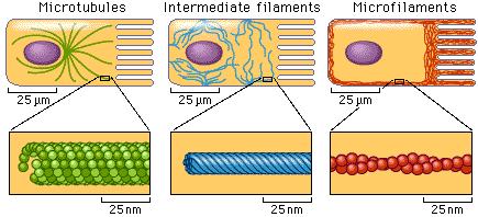
CYTOSKELETON
Cytosol contains a cytoskeletal fabric formed of microtubules, microfilaments, and intermediate filaments.
COMPOSITION:
The main proteins forming the cytoskeleton are tubulin, actin, myosin, tropomyosin and others which are also found in muscles.
MICROTUBULES INTERMEDIATE FILAMENTS MICROFILAMENTTS
MICROTUBULES:
- Microtubules are long, unbranched, slender tubulin protein structures.
- Microtubules provide support and shape to the cell and cellular membranes.
- Microtubules are involved in transportation of organelles within the cell and act as conveyor belts.
- Aggregation of microtubules give rise to various cell structures such as cilia, flagella, and centriole.
- The most important function of microtubules is their role in the assembly and dis-assembly of the spindle structure during mitosis.
MICROFILAMENTS:
- Microfilaments are slender cylindrical structures made up of contractile actin protein, linked to the inner surface of plasma membrane.
- Microfilaments are involved in internal cell motion
INTERMEDIATE FILAMENTS:
- Intermediate filaments have a diameter in between that of microtubules and microfilaments.
- The play a role in the maintenance of cell shape.
FUNCTIONS OF CYTOSKELETON:
- Several organelles are derived from special assemblies of microtubules such as cilia, flagella, basal bodies and centrioles.
- Movements such as cyclosis (cytoplasmic streaming movement) and ameboid movements are a function of microfilaments.
- Intermediate filaments are responsible for the determination of cell shape and integration of cellular compartments.
CENTRIOLE
Centrioles are organelles resulting from the aggregation of microtubules.
OCCURRENCE:
- Centrioles are present in Animal cells, cells of some micro-organisms and lower plants.
- Centrioles are absent in higher plants.
LOCATION: Centrioles mostly occur in a pair and are located near the exterior surface of the nucleus.
STRUCTURE:
- The cross-section of a centriole reveals cylindrical arrangement of 9 microtubule triplets (arranged into a ring like form).
- One microtubular triplet consists of one complete microtubule and two partial microtubules.
CENTROSOME:
The arrangement of two centrioles at right angles to each other gives rise to a centrosome.
FUNCTION OF CENTRIOLE:
- Centrioles play an important role in the location of furrowing during cell division.
- Just before cell division, the centrioles duplicate and each pair migrates to the opposite side of the nucleus. The spindle then forms between them.
- Centrioles also play an important role in the formation of cilia and flagella. It acts as a base from which the microtubular core of cilia and flagella arise.
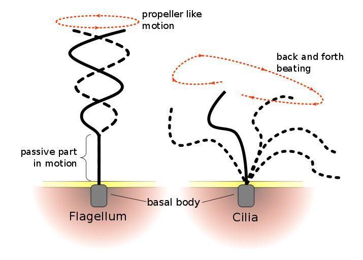
CILIA
Cilia are slender, hair like protuberances extending from the surface of eukaryotic cells.
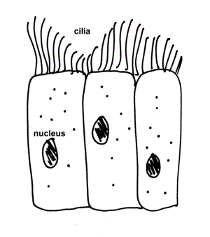
LOCATION: Cilia maybe found in the cells of the lungs, respiratory tract, middle ear, kidney tubules etc.
STRUCTURE:
- Cilia are mainly composed of microtubules.
- These microtubules arise from centrioles present immediately beneath the cell membrane and are known as basal bodies.
- An axoneme is the microtubular cytoskeletal structure that forms the core of a cilium.
- An axoneme has microtubular pairs arranged around central single microtubules.
- The axoneme structure is different for both types of cilia.
TYPES OF CILIA:
Cilia are of two types:
- Motile cilia
- Non-motile/ primary cilia
FLAGELLA
Flagella, a helical structure, is the extension of cell membrane responsible for the movement of associated cell.
STRUCTURE:
- Flagella has the same structure as that of motile cilia.
- Axoneme forms the main core of the structure.
- Axoneme consists of nine microtubule pairs arranged around two central single microtubules form the core of the flagella (9+0 arrangement).
- The microtubules arise from basal body which is a centriole present immediately beneath the cell membrane.
- The entire flagella is covered by extension of the cell membrane.
FUNCTION:
- Flagella is responsible for the locomotion of the cell.
- Flagella also act as a sensory organ which can detect changes in pH and temperature.
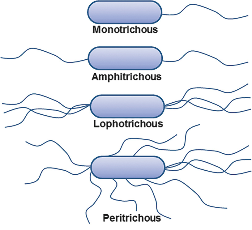
TYPES OF FLAGELLA:
There are five types of flagella based on their arrangement.
- Monotrichous: A single flagellum at one end of the cell.
- Amphitrichous: occurrence of two flagella, one at each end of the cell.
- Lophotrichous: multiple flagella at one end of the cell.
- Peritrichous: Several flagella distributed over the entire surface of cell.
DIFFERENCE BETWEEN FLAGELLA AND MOTILE CILIA:
| FLAGELLA | CILIA |
| Flagella are longer in length | cilia are shorter in length |
| Flagella are fewer in number | Cilia are present in large number |
| Flagella are usually present at one end of the cell | Cilia are usually present over the entire surface of the cell |
| Flagella exhibit coiled motion | Cilia exhibit radical motion |
| Flagella are present in both prokaryotes and eukaryotes | Cilia are only found in eukaryotes |
MITOCHONDRIA
Mitochondria is a membrane bound organelle often termed as the powerhouse of the cell.
STRUCTURE:
- Mitochondria is a rod/ sausage shaped double membranous structure with an outer membrane and inner membrane separated by a small space.
- The outer membrane is smooth and consists of transport proteins called porin which forms aqueous channels allowing access to water soluble molecules from the cytoplasm.
- The space between the outer an inner membrane is called inter-membrane space.
- The inner membrane is thrown into folds and surrounds the mitochondrial matrix.
- The infoldings of inner membrane form finger-like projections into the matrix called cristae, the purpose of which is to increase the surface area for ATP production.
- The inner membrane is far more selective and only allows passage of materials via specialized transport proteins.
- the mitochondrial matrix is enclosed by the inner membrane and includes various enzymes, co-enzymes, organic and inorganic salts involved in the metabolic processes of mitochondria.
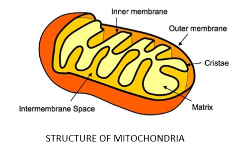
FUNCTIONS:
- mitochondria carry out various metabolic processes like Krebs’s cycle, aerobic respiration, fatty acid metabolism etc.
- as a result of these processes, energy is extracted from the organic compounds and is packed into energy rich ATP molecules.
- The ATP is then utilized by the cell for provision of energy on demand.
PLASTIDS
Plastids are membrane bound, pigment containing organelles which are mainly present in plant cells only.
Plastids consists of three types namely:
- Chloroplasts
- Chromoplasts
- leucoplasts
CHLOROPLAST:
Chloroplast are green pigment (chlorophyll) containing plastids found mostly in photosynthetic plants.
CHLOROPHYLL:
- Chlorophyll, the green pigment, is the organic compound responsible for helping the cell to absorb light energy and utilize it to manufacture food.
- Chlorophyll structurally resembles the haem group of haemoglobin, a protein used in the transport of oxygen
- The main difference between these two molecules is that chlorophyll has Mg++ while haem has Fe++ as central atom.
STRUCTURE OF CHLOROPLAST:
A chloroplast consists of three main structural components
- Envelope
- Stroma
- Thylakoid
Envelope:
It is a double membranous structure enclosing the stroma:
Stroma:
- It is the fluid which surrounds the thylakoids.
- It contains proteins, some ribosomes and a small circular DNA
- It is in this part of the chloroplast where CO2 is fixed to manufacture sugars.
- Some proteins are also synthesized in the stroma.
Thylakoid:
- Thylakoids are flattened vesicles which arrange in stacks to form grana and intergrana.
- A granum appears to be a pile of thylakoids stacked in the form of coins.
- On an average, there are 50 or more thylakoids piled to form one granum.
- Chlorophyll molecules are embedded in the grana membrane and therefore grana membrane serves as a site for trapping light energy and generating ATP.
CHROMOPLAST:
- Chromoplast contains yellow an orange pigment an are found in the petals of flowers and in the ripened fruit.
- Chromoplast help in pollination and dispersal of seeds.
LEUCOPLAST:
- Leucoplasts are colourless and triangular/tubular in shape.
- They are present in the underground parts of plants and store food.
ENDOSYMBIONT THEORY:
Endo means inside and symbiont is an organism which lives in a mutually beneficial relationship with another organism.
Endosymbiont theory determines mitochondria and chloroplast to be ancient bacteria that now live inside larger eukaryotic cells.

Responses